Topic 3 | Proteins and Structures
📅 Date: 25 September 2025
📖 Synopsis: Exploring databases: UniProt, RCSB PDB
Lecture topics
- Accessing protein annotation, domains, and sequence features - UniProt
- Visualising receptor structures - RCSB PDB
- Exploring ligand binding sites in protein complexes - RCSB PDB / UniProt
Learning goals
- Learn to retrieve and interpret protein functional information.
- Explore 3D receptor structures.
- Integrate protein sequence and structural data.
Introduction
This class explores two complementary databases: UniProt and RCSB PDB.
UniProt focuses on proteins—their sequences, functions, domains, isoforms, variants, and biological context, while RCSB PDB is the central archive of experimentally determined 3D macromolecular structures and related ligands.
Together, they show how sequence-level knowledge and structure-level evidence combine to explain protein function and drug interactions. Learning to navigate both is essential for connecting what a protein is (UniProt) with how it looks and works in 3D (PDB).
Challenges
Using these resources for the first time presents common difficulties:
- Information overload: both UniProt and PDB are rich and dense; it takes practice to identify what is essential for your question.
- Identifier mapping: connecting a UniProt accession to the correct PDB structure(s) can be tricky (isoforms, chain IDs, residue numbering).
- Quality and interpretation: structures vary by method (X-ray, cryo-EM, NMR), resolution, and completeness; understanding validation metrics is important.
- Navigation structure: UniProt integrates many cross-references (InterPro, Pfam, Reactome, KEGG, etc.); PDB integrates tools, visualisers, and related databases (EMDB, PDBe-KB).
- Technical terminology: domains, motifs, PTMs, variants, assemblies, interfaces, and binding sites may be unfamiliar at first.
- Search skills: choosing effective queries (gene symbol vs UniProt accession; PDB ID vs ligand name) and refining filters is key.
As always: It’s normal to feel a bit overwhelmed at the start. These databases are extensive, and nobody masters them immediately. With practice, their structure becomes familiar and you will quickly find what you need. Do not worry if it feels slow initially—each search builds confidence, and over time the process becomes natural. By the end of these classes you will feel confident navigating core protein and structure databases.
Databases overview
UniProt
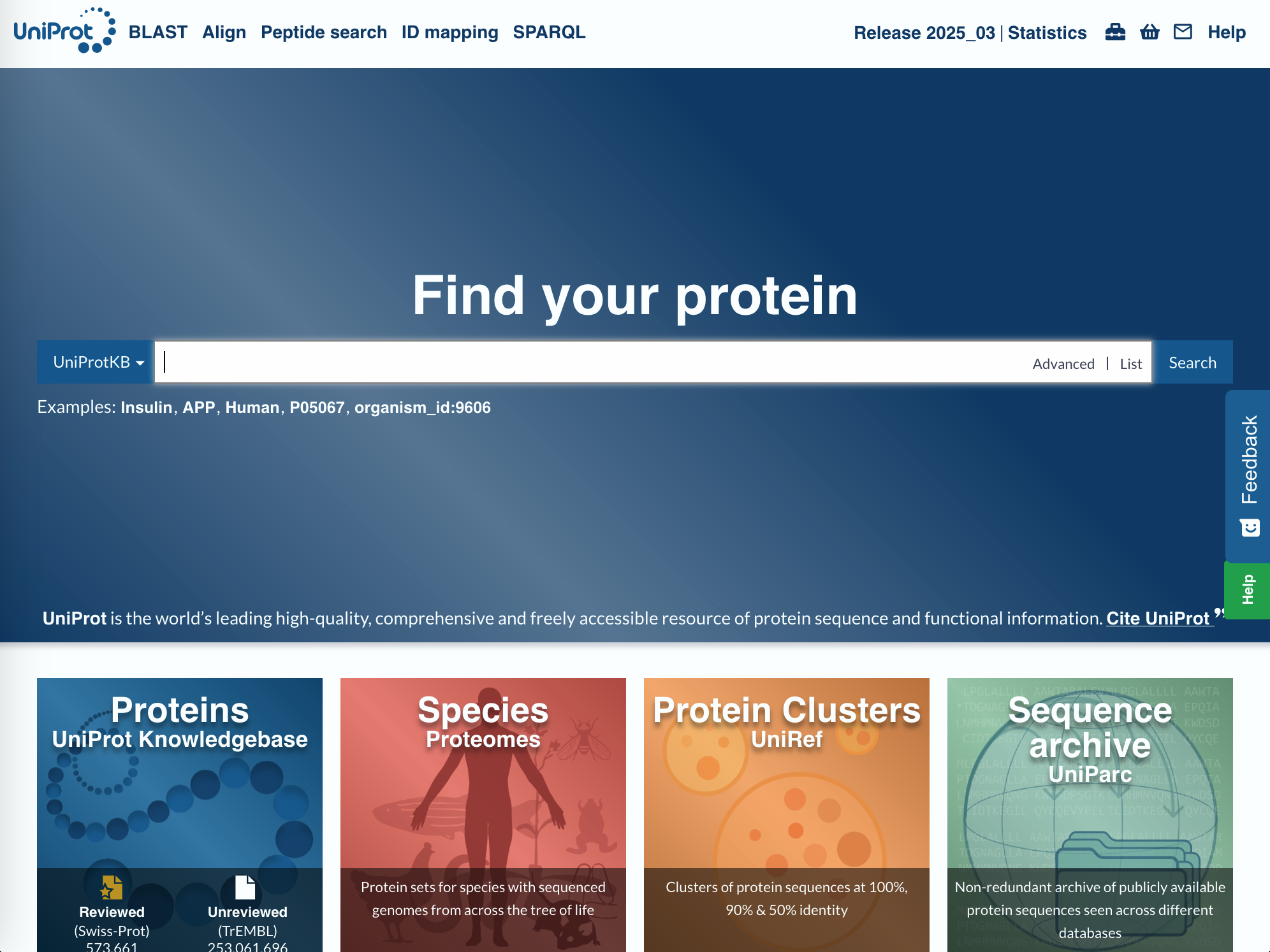
1. Purpose | A comprehensive knowledgebase of protein sequences and functional annotations.
2. Content | Protein names, gene symbols, isoforms, domains/motifs, post-translational modifications (PTMs), subcellular localisation, interaction partners, variants/disease associations, pathways, cross-references, and evidence codes.
3. Typical Use Cases
- Retrieve a canonical protein sequence and known isoforms.
- Identify functional domains/motifs and PTMs relevant to activity or regulation.
- Review variants (position, effect, disease links) and map them to the sequence.
- Find pathway context (via Reactome/KEGG) and protein–protein interactions.
- Jump to 3D structures and structure coverage via cross-links to PDB.
4. External Connections | Cross-linked with RCSB PDB (structures), NCBI, Ensembl, InterPro (domains), Reactome/KEGG (pathways), ClinVar/OMIM (clinical), ChEMBL/DrugBank (bioactive compounds/targets), among others.
RCSB PDB – Protein Data Bank
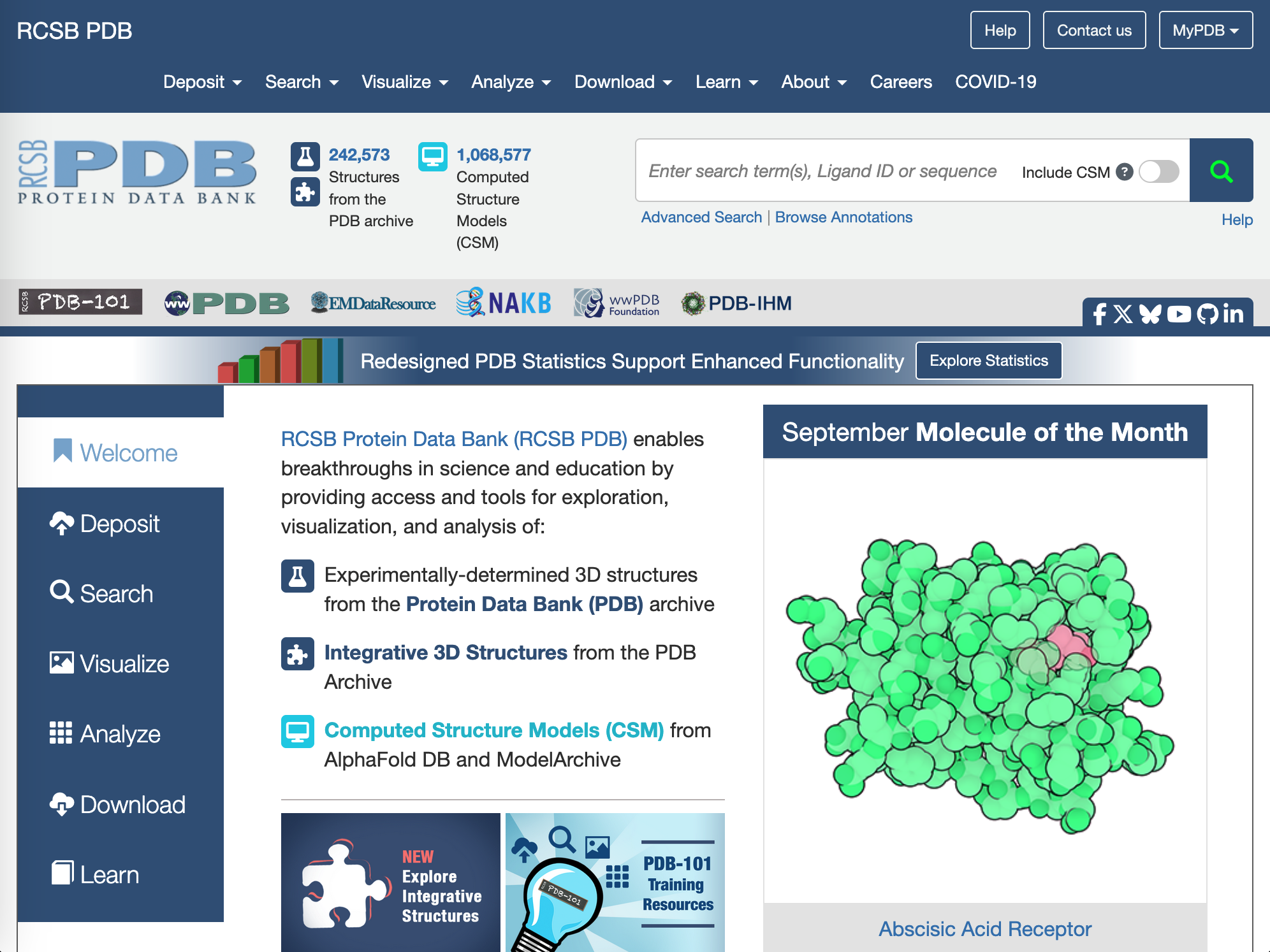
URL: https://www.rcsb.org
1. Purpose | The primary public archive of experimentally determined 3D structures of proteins, nucleic acids, and complexes, with tools for visualisation and analysis.
2. Content | Atomic coordinates, experimental method (X-ray, cryo-EM, NMR), resolution/validation metrics, biological assemblies, ligands/cofactors, binding sites, sequence-to-structure mappings, and annotations on interfaces and dynamics.
3. Typical Use Cases
- Find PDB structures for a protein target and inspect ligand binding or active sites.
- Compare alternative structures (different ligands, conformations, mutants, species).
- Assess model quality (resolution, R-factors, MolProbity/validation reports).
- Map sequence positions (variants, PTMs, domain boundaries) onto 3D coordinates.
- Download structures for molecular graphics or downstream analysis.
4. External Connections | Integrated with UniProt (sequence mapping via SIFTS), PubChem/ChEMBL (ligands), NCBI/Ensembl (gene/protein), Reactome/KEGG (pathways), and literature via PubMed.
TP Activity 3
Diazepam & GABA Receptor Structures
Goal
How to work
- Search for diazepam in DrugBank.
- Open the DrugBank entry and follow the link to the UniProt page of diazepam’s molecular target.
- In UniProt, explore the entry to find information about the protein sequence, function, and biological relevance. Use the worksheet as a guide.
- From UniProt, follow the links to RCSB PDB to find available 3D structures of the protein, and examine how these structures help explain its function and drug interactions.
Evaluation
- Select 4 facts from each database that were surprising or new to you.
- For each fact, briefly explain why it was surprising or new.
- Write your findings in a Word document and submit it in the Tutoria before the end of class.
Hints
- UniProt entries link directly to PDB and other databases. Focus on information that helps you understand how diazepam interacts with its molecular target.
- Take your time to understand the type of information each database provides and be critical of the info shown.
- Ask the tutor if you have any questions.
- Discuss with your colleagues how they interpret the different sources of information, and compare with your ideas.
- Keep in mind: always use your critical thinking.